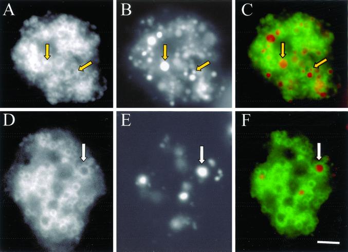
Figure 9.
GFP-Rab7 localizes to macropinosomes, lysosomes, and postlysosomes. Cells expressing GFP-Rab7 were allowed to attach to coverslips and were pulsed with Texas Red dextran for 10 min followed by a 60 min chase in marker-free medium to load lysosomes and postlysosomes (A, GFP; B, Texas Red; C, merge), or cells were pulsed for 5 min with Texas Red dextran to load macropinosomes (D, GFP; E, Texas Red; F, merge). Cells were then fixed with 1% formaldehyde in HL5, and images were captured with the use of a fluorescence microscope equipped with a charge-coupled device camera. Arrows in A, B, and C denote a postlysosome and a lysosome from left to right, and arrows in D, E, and F denote a macropinosome. Bar, 10 μm.

