FIG 2.
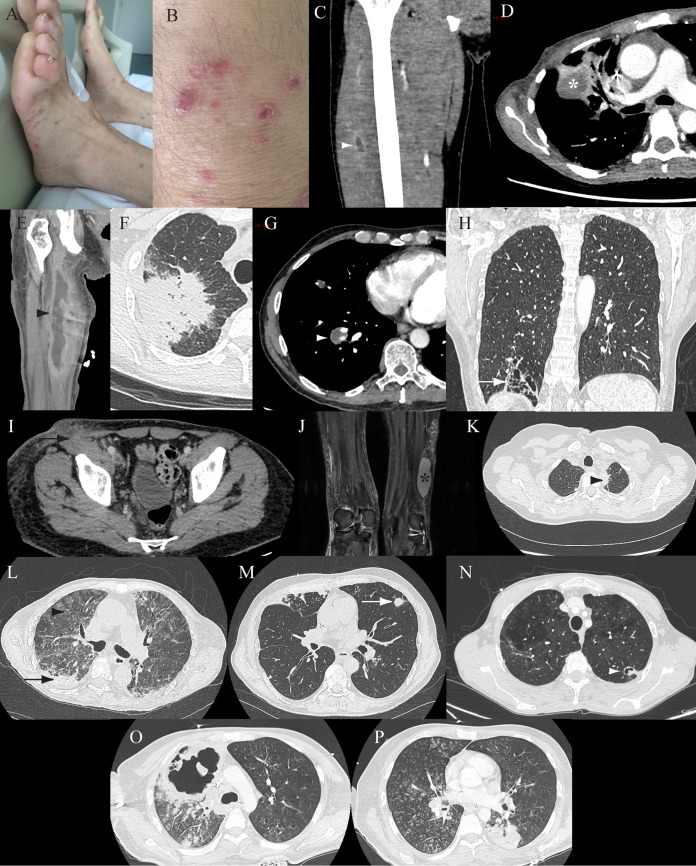
Clinical and radiological features in a series of 13 patients with probable nocardiosis. (A and B) Patient 1, multiple superficial and purple nodular skin lesions on both legs. (C) Patient 2, coronal enhanced computed tomography (CT) of the right leg, showing a muscle abscess of the thigh (white arrowhead, central low attenuation with peripheral enhancement). (D and E) Patient 3, axial enhanced lung CT (D), showing a right pulmonary mass (white star, central attenuation), and coronal enhanced CT of the right leg (E), showing a muscle abscess of the thigh (black arrowhead, central attenuation with peripheral enhancement). (F) Patient 4, axial nonenhanced lung CT, showing a right spiculated lung consolidation. (G) Patient 5, axial enhanced lung CT, showing a right pulmonary mass (white arrowhead, low density). (H) Patient 6, coronal enhanced lung CT, showing localized bronchiectasis with thin surrounding lung consolidation (white arrow). (I) Patient 7, axial enhanced abdominal CT, showing a subcutaneous abscess and lymph node involvement (black arrow). (J) Patient 8, coronal T2 fat-saturated magnetic resonance imaging of the legs, showing a subcutaneous abscess (black star, hyperintensity). (K) Patient 9, axial nonenhanced lung CT, showing a left apical lung mass (black arrowhead). (L) Patient 10, axial enhanced lung CT, showing a lung consolidation (black arrow) and diffuse ground-glass opacities (black arrowhead). (M) Patient 11, axial nonenhanced lung CT, showing a lung nodule (white arrow). (N) Patient 12, axial nonenhanced lung CT, showing a cavitated lung nodule (white arrowhead). (O and P) Patient 13, axial nonenhanced lung CT, showing bilateral involvement with excavation of 10 cm, micronodules, and lung consolidation.

