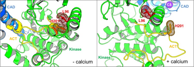
FIG 3.
Location of amino acids mutated in CDPK1 in clones A-f8 (H201Q) and C-d12 (L96P). The images are close-ups of the region of CDPK1 where mutations are located. The kinase domain is in green, with its activation segment (ACT) in yellow, and the CAD-EF hand domain is in blue. The locations of the mutations are shown as stick residues (WT residues are shown) along with their van der Waals surfaces, based on structures in the presence or absence of calcium. The PDB accession number for the active form (with calcium) is 3HX4, and the PDB accession number for the inactive form (without calcium) is 3SX4.

