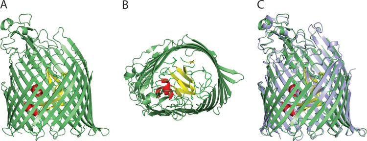
FIG 3.
Crystal structure of PiuD from P. aeruginosa. Side (A) and extracellular (B) views of PiuD. The 22-stranded transmembrane β-barrel is colored in green. β-Sheets of the plug domain are colored in yellow, loops in green, and helices in red. (C) Structural comparison between PiuA (light blue) and PiuD (green).

