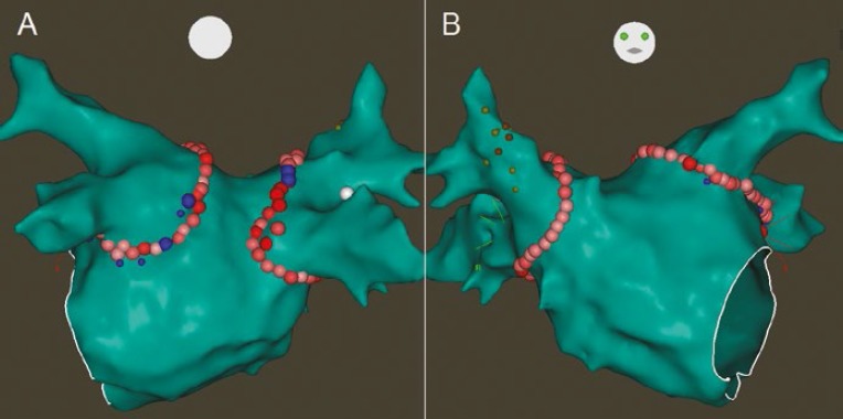
Figure 1: Ablation Lesion Set for Circumferential Pulmonary Vein Isolation.

Posterior (A) and anterior (B) projections of the left atrium on the 3-dimensional electroanatomical map showing circular ablation lesions delivered around both sets of pulmonary veins (pink, red and blue circles). The smaller yellow and orange circles in (B) are sites where pacing resulted in diaphragmatic stimulation, delineating the course of the right phrenic nerve.
