Figure 3.
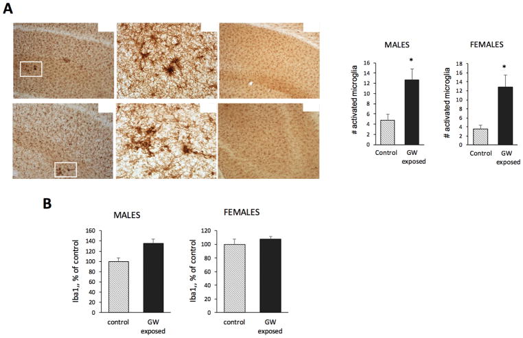
Microglial activation in the hippocampus of GW-exposed mice. A. Representative microphotographs showing that a larger number of activated microglia, prominent by their distinctive cell morphology and increased levels of Iba1 expression, were detected in the CA1-3 regions of the hippocampus of exposed CHGFP mice compared to controls in both female and male mice. Original pictures were taken at 100x and 400x, as indicated. The inset graphs are the results from the quantitation of the number of activated microglia in the CA1-3 region showing that in this region of the hippocampus the number of activated microglia is significantly larger in exposed female and males than in their respective control mice. B. Western blot analysis of protein extracts from the whole hippocampus with Iba1 antibodies. The level of Iba1 increased in exposed males and females to 135.5% and 107.8% of controls, respectively. Two-way ANOVA showed a significant effect of exposure for Iba1 (F = 4.63; p = 0.038), and no effect of sex.

