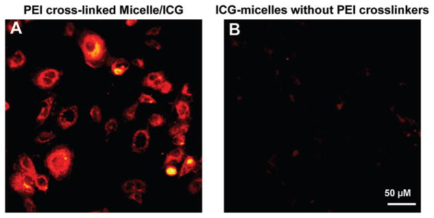
Figure 7.
ICG fluorescence images of A431 cells after incubation with PEI-cross-linked ICG nanocapsules (A) and ICG nanocapsules without PEI cross-linkers (B) for 30 min with an ICG concentration of 5 μg/mL. After washing with PBS three times, fluorescence microscopy images of the cells were acquired by a microscope (Zeiss Axiovert 200) with an ICG filter set (49030 ET, Chroma Technology Corp., excitation 775/50 nm, emission 845/55 nm) and a 20× objective (Zeiss, Ecplan-NEOFLUAR). Images were recorded using an EMCCD camera (Andor) with a gain of 200 and an exposure time of 0.2 s.

