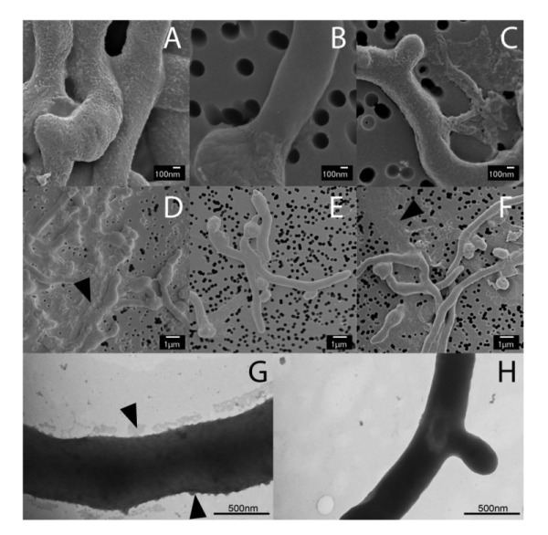
Figure 1. FIGURE 1: Electron micrographs revealing at Mat-dependent extracellular matrix.

Scanning electron micrographs of young vegetative mycelium (A-F) show an abundance of extracellular material covering the hyphae of wild-type S. lividans (A) and between hyphae (D, indicated by arrow). This extracellular material was also present in the cslA null mutant (C and F, indicated by arrow), but was absent in matAB null mutants (B and E). Negatively stained hyphae with tungsten acid, specific for polymeric substances, revealed a scabrous outside coating in wild-type hyphae (G) that is absent in the mat mutant (H). All strains were grown for 8 h in TSBS media in shake flasks.
