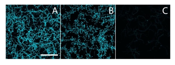
Figure 2. FIGURE 2: Calcofluor white staining to identify β-(1,4)-glycans.

S. lividans (A), and its matAB (B) and cslA (C) mutants were stained with calcofluor white (CFW) to identify the presence of extracellular β-(1,4)-glycans. The staining patterns indicated the presence of β-(1,4)-glycans in both the parental strain and its matAB mutant, but not in the cslA mutant. Bar, 50 µM.
