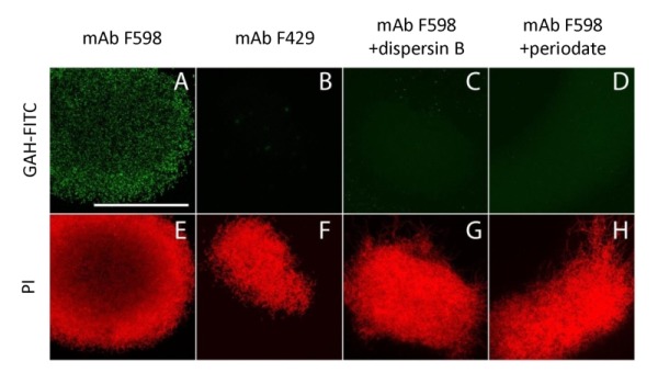
Figure 3. FIGURE 3: Immunofluorescence micrographs of S. lividans identifying extracellular PNAG.

Mycelia from 18 h old cultures of S. lividans 66 were analyzed for the presence of PNAG with the monoclonal antibody mAb F598 and secondary anti-human IgG Alexa 488 conjugate (green) (A,C,D). As controls for the specificity of the primary antibody we used mAb F429, a monoclonal antibody that binds alginate (B), and samples treated with 50 µg/ml dispersin B (C) that degrades PNAG or with 0.4 M periodate (D), which degrades β-(1,6)-glycans. To visualize the hyphae, the DNA was stained with propidium iodide (red) (E-H). Bar, 100 µm.
