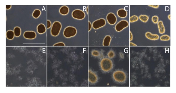
Figure 5. FIGURE 5: Effect of cellulases and dispersin B on mycelial morphology.

Light micrographs show S. lividans 66 (A), S. lividans 66 treated with 2 U/ml cellulase (B), 100 µg/ml dispersin B (C) or both cellulase and dispersin B (D), the matB (E) and cslA mutants (F) and the cslA mutant harboring pMAT7 without (G) or with (H) added dispersin B. All strains were grown in TSBS medium for 24 h at 30oC. The effects on morphology were visualized by wide-field microscopy. Bar, 500 µm.
