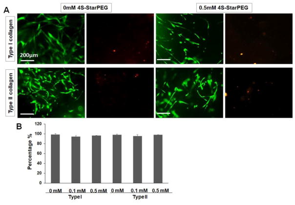
Figure 1.
LIVE/DEAD® cell viability assay for DPSCs grown in collagen hydrogels: (A) Most cells grown in non-crosslinked and 4S-StarPEG crosslinked collagen hydrogels exhibiting high cell viability. Scale bar: 200 μm. The images of same field were taken to show the green or red fluorescence – labeled cells. Live cells labeled with calcein AM (green). Dead cells labeled with ethidium homodimer-1 (red). (B) Percentage of live cells in collagen hydrogels as determined by LIVE/DEAD® cell assay.

