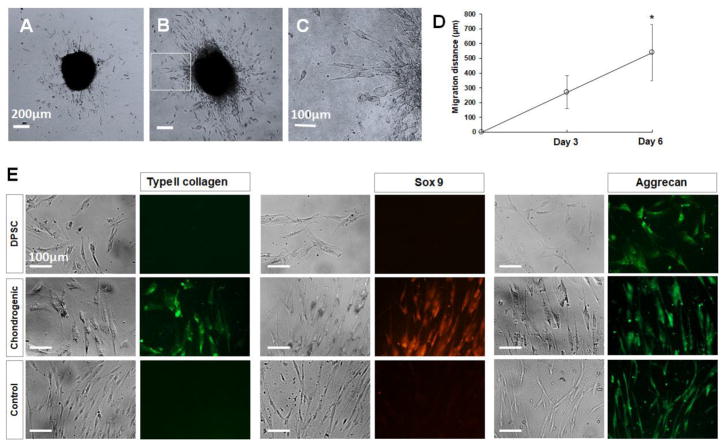
Figure 4.
Migration and differentiation of DPSC-derived chondrogenic cells. (A–D) DPSC-derived chondrogenic cells migrated out of cell pellet after culturing on collagen-coated cell culture dish. Scale bar Figures A and B: 200 μm. Scale bar Figure C: 100 μm. (E) Cells that migrated out of the pellets labeled with anti-type II collagen, anti-sox 9, and anti-aggrecan antibodies. Scale bar: 100 μm.

