
Fig 3. Schematic drawing and anatomical demonstration of the GG injection site.
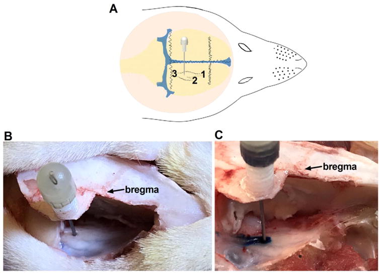
(A) Schematic drawing showing the site of the GG injection. The structure details: 1) V1/V2 (ophthalmic/maxillary), 2) V3 (mandibular), and 3) sensory root. (B) Anatomical position of the cannulation. (C) Methylene blue injection showing the drug injection site.