
Fig 6. HCN1 and HCN2 immunostaining of the GG sections.
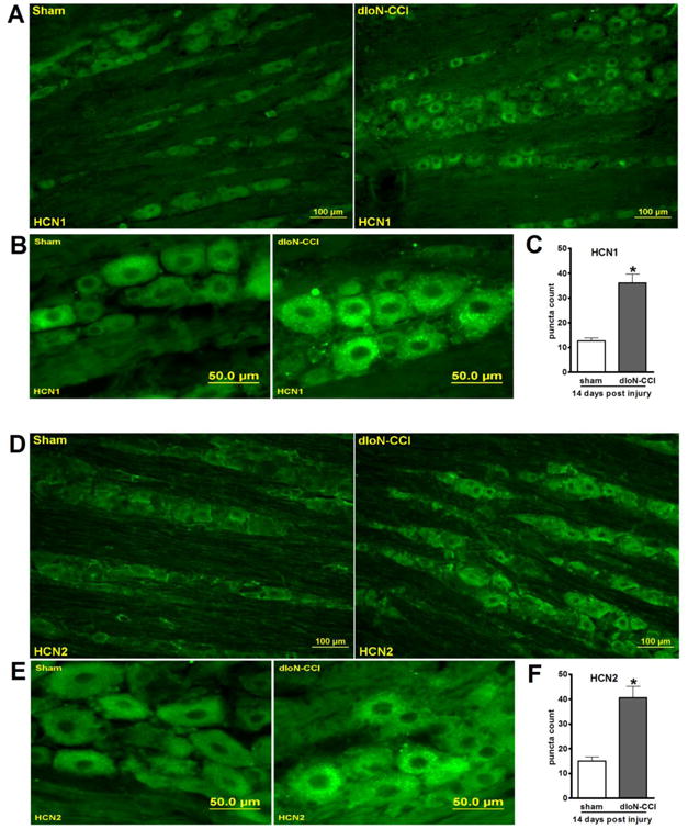
Representative micrographs of the ipsilateral GG isolated from sham or dIoN-CCI (14 days) rats were stained with anti-HCN1 (A-B) or anti-HCN2 (D-E) antibody. The ipsilateral GG of dIoN-CCI rat showed increase in HCN1 (C) and HCN2 (F) immuno-positive puncta counts. *p<0.001, n=4