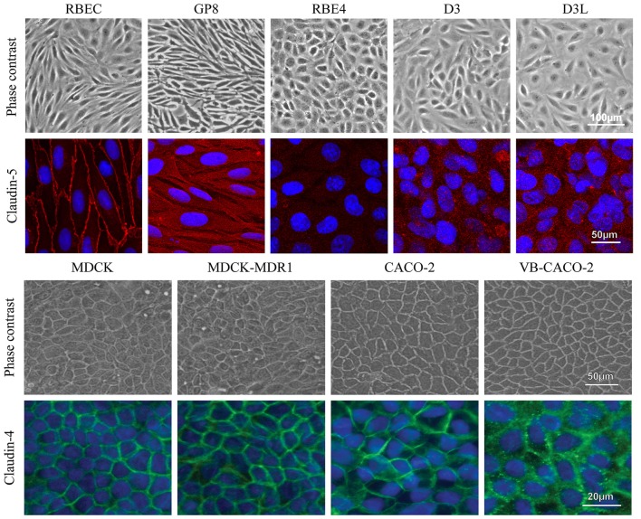
Figure 9.
Different types of brain endothelial and epithelial cell cultures examined by phase contrast microscopy and stained for tight junction proteins CLDN5 (red) and CLDN4 (green), and cell nucleus (blue). The CLDN5 immunostaining was well visible on the cell border of RBEC cells, while it was very weak or undetectable in the brain endothelial cell lines. MDCK and Caco-2 epithelial cells showed an intense staining for CLDN4. Bars: 50 and 100 μm (phase contrast pictures); 20 and 50 μm (immunostaining pictures).

