Figure 2.
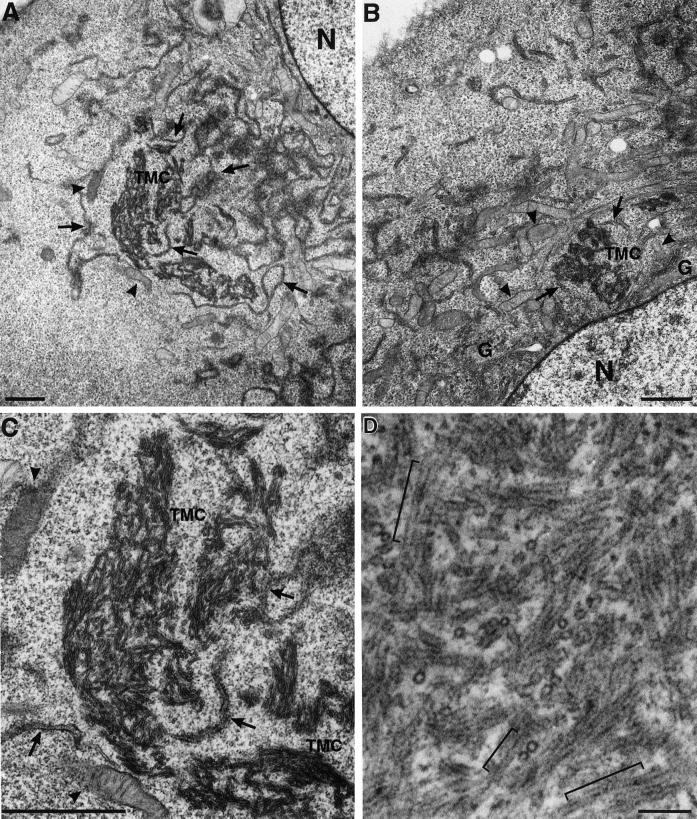
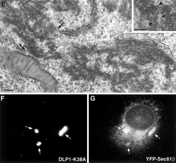
DLP1-K38A aggregates are comprised of constricted ER membrane tubules with periodic DLP1 striations. A plasmid encoding GFP-DLP1-K38A was microinjected into nuclei of BHK-21 (A–C) or Clone 9 (D) cells. 24 h after injection, cells were fixed, embedded, and subjected to thin-section electron microscopy. The peripheral cytoplasm of these cells display little ER or mitochondria. Instead, multiple TMCs are found in proximity to an atrophied endoplasmic reticulum (A–C, arrows) or collapsed mitochondria (A–C, arrowheads) near the nucleus. Higher magnification of a TMC from A reveals a complex network of membrane tubules (C) that exhibit a consistent uniform diameter. TMCs exhibit dense periodic striations or collars along the length of tubules (D). Higher magnification TEM image shows lipid bilayers in tubules (E). Although lipid bilayers are apparent in most tubules, they are especially prominent in the bracketed region in E and in cross sections in inset marked by arrowheads. Arrows in E point to the apparent connection between the TMC and ER tubules. (F and G) TMCs contain the ER transmembrane protein Sec61β. BHK-21 cells stably expressing YFP-Sec61β were transfected to induce the formation of TMCs and stained with anti-DLP1 antibodies. TMCs shown in F are positive for YFP-Sec61β (G), indicating TMCs are comprised, in part, of ER membrane. N, nucleus; G, Golgi apparatus. Bars, 1.0 μm (A–C), 100 nm (D and E)..


