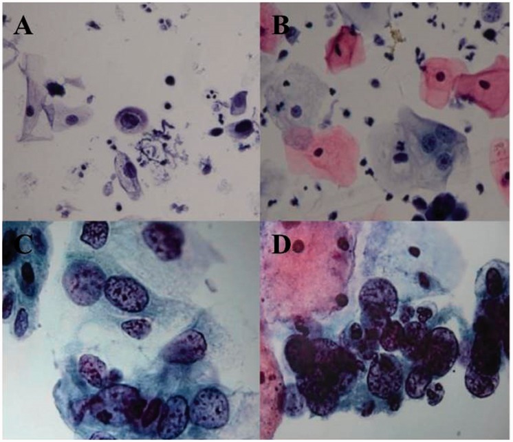
Figure 2.
Representative photomicrographs of the typical appearance of four subtypes of cervical epithelial lesions as they appear during the ThinPrep cytological test: (A) atypical squamous cells with undetermined significance; (B) low-grade squamous intraepithelial lesion; (C) high-grade squamous intraepithelial lesion; (D) squamous cell carcinoma. The colour version of this figure is available at: http://imr.sagepub.com. Scale bar 10 µm.

