
Fig. 2.
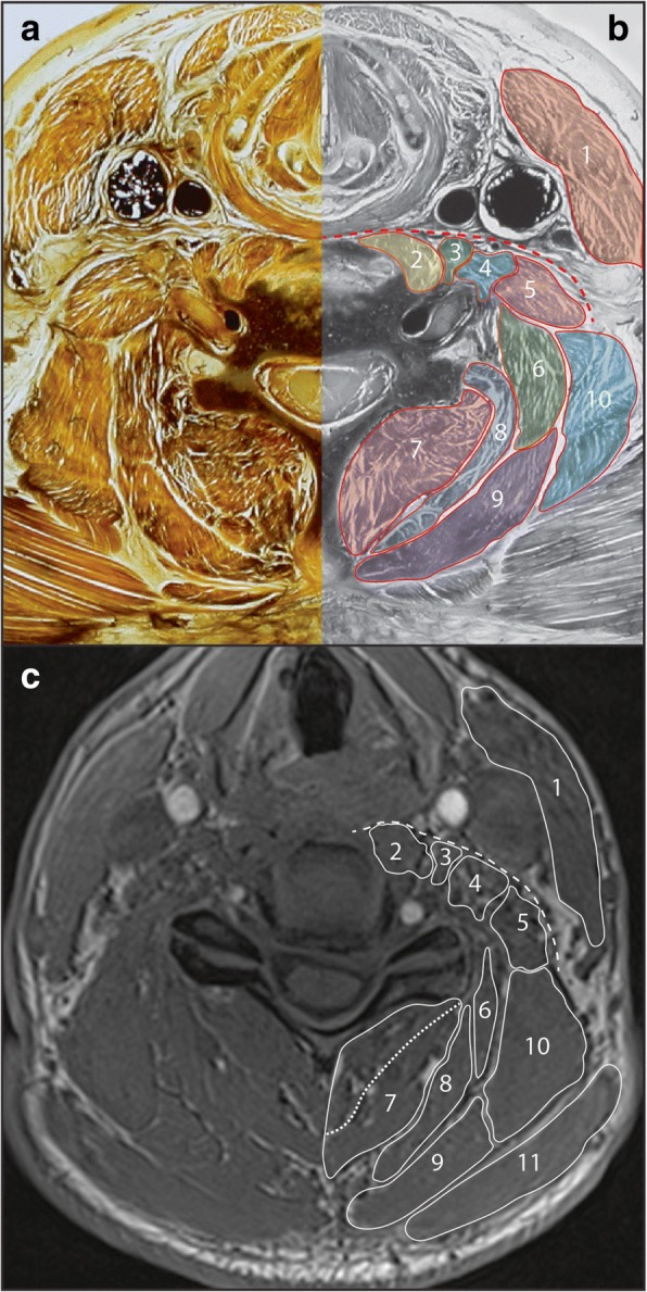
Axial E12 plastinated section (a) with schematic illustration (b) and in-phase magnetic resonance image (c) at approximately C5/6 identifying musculature at this vertebral level. Dashed red (b) and white (c) line indicates an anatomical plane which can be used as a reference point for identifying some anterior muscles. Dashed white line in (c) indicates likely border between multifidus and semispinalis cervicis. 1. Sternocleidomastoid; 2. Longus colli; 3. Longus capitis; 4. Scalenus anterior; 5. Scalenus medius; 6. Splenius cervicis; 7. Multifidus / semispinalis cervicis; 8. Semispinalis capitis; 9. Splenius capitis; 10. Levator scapulae; 11. Trapezius