Figure 3.
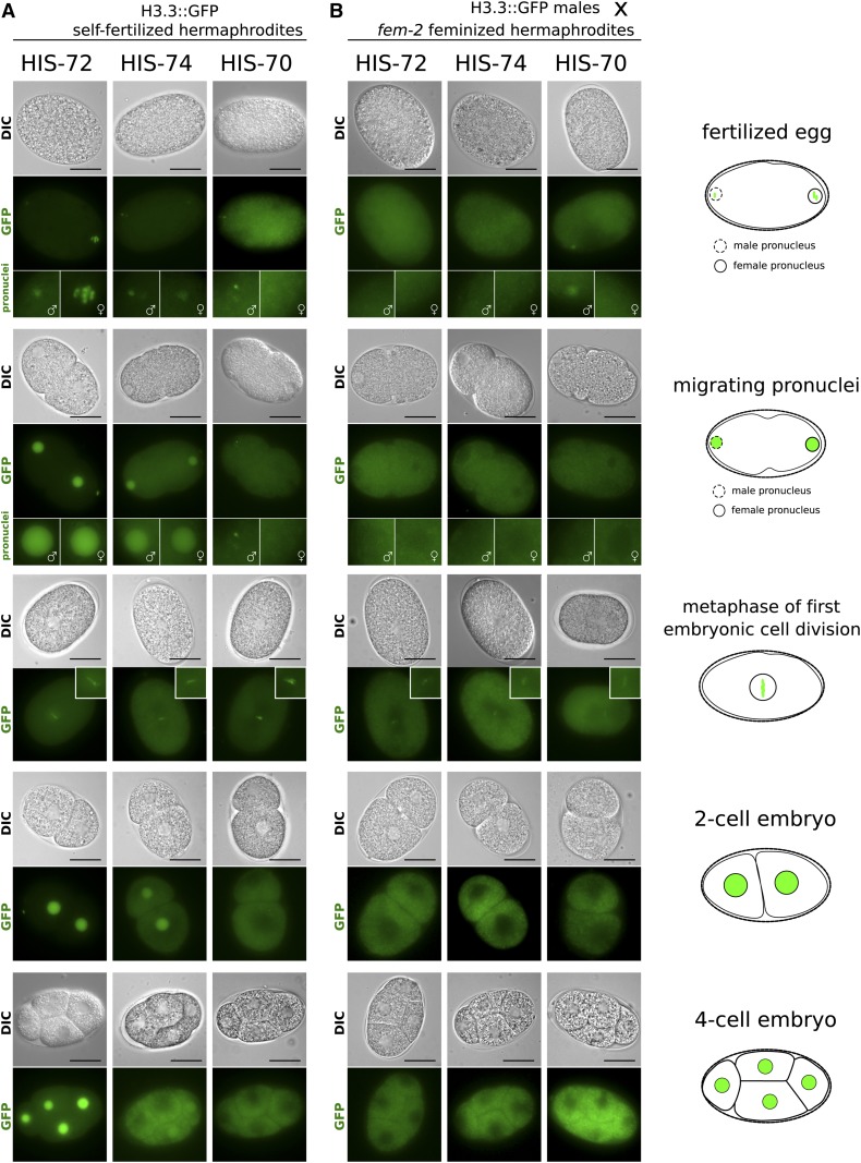
Dynamics of H3.3 proteins in early embryos. Representative live-cell images of early stages in embryonic development are shown. Stages include eggs after fertilization, migrating pronuclei, the metaphase of first cell division, the two-cell embryo, and the four-cell embryo, as outlined by cartoons on the right. (A) Eggs of self-fertilizing hermaphrodites carrying H3.3::GFP fusions. HIS-72 (left panels) is present in both male and female pronuclei upon fertilization and remains chromatin associated during metaphase of the first mitotic division. It is subsequently present in all cells. HIS-74 (center panels) is present in both male and female pronuclei upon fertilization and remains chromatin associated during metaphase of the first mitotic division. It is present in both nuclei at the two-cell stage, but subsequently becomes diluted. It is sometimes still faintly visible in two nuclei at the four-cell stage and then becomes undetectable. HIS-70 (right panels) is faintly detectable in the male but not the female pronucleus, remains chromatin associated during the first mitotic division, and subsequently becomes undetectable. (B) Eggs of feminized fem-2 hermaphrodites fertilized by males carrying H3.3::GFP fusions. HIS-72 and HIS-74 are not detectable in the pronuclei, while HIS-70 is visible in the male pronucleus. All three GFP fusions appear faintly chromatin associated during the first mitotic division and become undetectable after that. In GFP images representing fertilized eggs and migrating pronuclei, female pronuclei are highlighted by enlarged images with female symbols and male pronuclei are highlighted by enlarged images with male symbols. Bars represent 20 µm.

