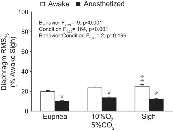
Fig. 4.

Bilateral average amplitude of the RMS EMG signal at 75 ms (RMS75) across behaviors during awake and anesthetized conditions. RMS75 values were normalized to the awake sigh peak RMS EMG for each hemidiaphragm within each animal (n = 9). A main effect of behavior and condition was observed. Post hoc comparisons: *P < 0.05 compared with awake RMS75 for the same behavior; ‡P < 0.05 compared with awake eupnea.
