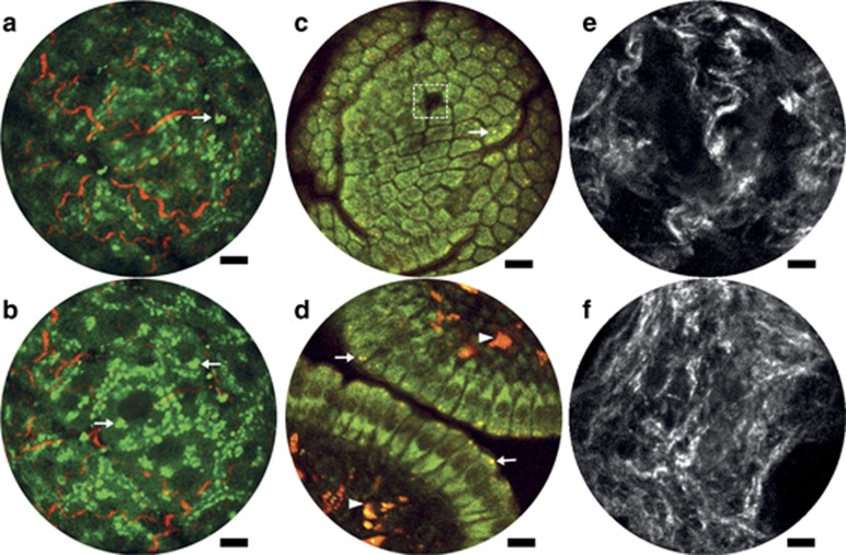
Figure 3.
Endomicroscopy 2PF and SHG label-free structural imaging. (a, b) Overlay of intrinsic 2PF and SHG images acquired from mouse liver ex vivo. The emission signal was detected through two spectral channels: 496–665 nm (green, 2PF signal) and 435–455 nm (red, SHG signal). (c, d) Two-photon autofluorescence images of the mucosa of mouse small intestine in vivo. The two detection channels are 417–477 nm for NADH (green) and 496–665 nm for FAD (red). (e, f) SHG images of cervical collagen fiber network acquired through intact ectocervical epithelium of cervices dissected from preterm-birth mouse models (e) and normal pregnant mice (f) at gestation day 15. All images were acquired using the 200-μm WD miniature objective. The excitation conditions include ~30 mW at 890 nm (a, b), ~30 mW at 750 nm (c, d) and ~40 mW at 890 nm (e, f). Four raw frames are averaged in a–d and 10 in e, f, corresponding to an effective frame acquisition time of ~1.5 s (a–d) and ~3.8 s (e, f), respectively. Scale bar=10 μm.

