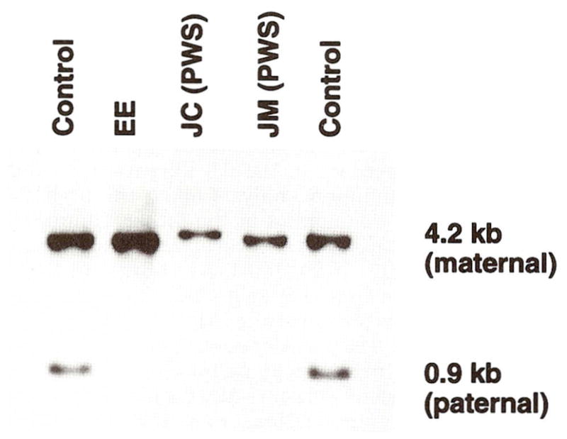
Fig. 5.

Southern hybridization using radio-labeled SNRPN CpG island probe (0.9 kb) [Sutcliffe et al., 1994] hybridized to 5 μg genomic DNA isolated from peripheral blood and digested with Xba I and Not I enzymes, produces two different size fragments (upper 4.2 kb band from the maternal chromosome 15 and lower 0.9 kb band from the paternal chromosome 15). Five individuals were studied. Our patient (EE) and two PWS patients (JC and JM) showed only the upper band (maternal) indicating the PWS diagnosis or absence of the lower band (paternal) while the two controls have both the upper and lower bands.
