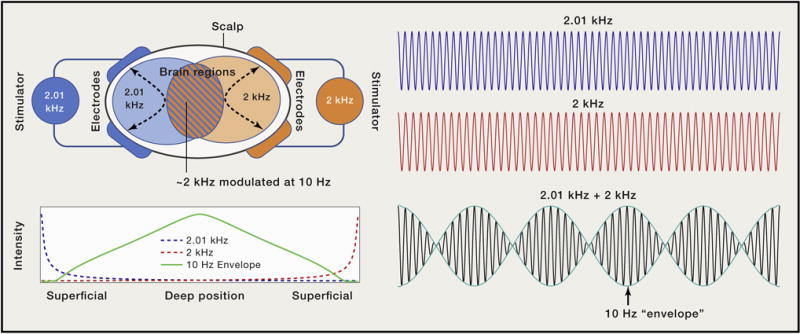
Figure 1. Temporal Interference for Noninvasive Electrical Stimulation of Deep Brain Regions.

Two pairs of electrodes on the surface of the head concurrently deliver electric currents oscillating at high but slightly different frequencies (e.g., 2 kHz, blue; 2.01 kHz, orange). In the brain regions near each pair, high-frequency electric fields of corresponding frequency and fixed amplitude are generated. Where the two frequencies overlap, the superposition of the two sinusoids produces a slowly “beating” envelope at a frequency equal to the difference of the two sinusoids (e.g., 10 Hz, blue/orange stripes). While the intensity of the individual high-frequency electric fields is maximal at superficial regions, the intensity of the slow beat frequency peaks in the region marked by equal contribution of both signals, which coincides in this case with the middle of the brain.
