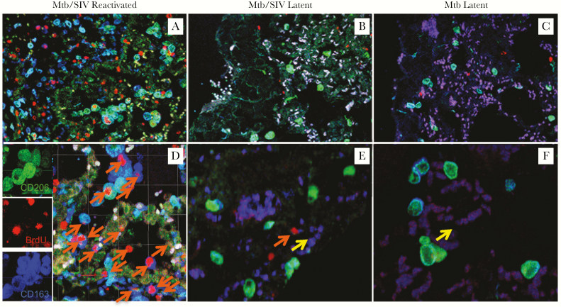
Figure 4.
Increased turnover of lung macrophages in the Mycobacterium tuberculosis (Mtb)/simian immunodeficiency virus (SIV)-coinfected rhesus macaques correlates with reactivation of latent tuberculosis (TB). (A–F) Confocal microscopy was used to analyze lung tissue sections stained with anti-CD206 (mature macrophages; green), anti-5-bromo-2’-deoxyuridine (BrdU) (turnover; red), and anti-CD163 (macrophages; blue), in Mtb/SIV-coinfected macaques with signs of TB reactivation (A and D, n = 2), Mtb/SIV-coinfected animals that remained latent (B and E, n = 2), as well as from the lungs of macaques infected only with Mtb that remained latent (C and F, n = 2). Images were captured from the capsid region of granuloma with a Leica TCS SP2 confocal microscope equipped with 3 lasers (Leica Microsystems) at ×200 (A, B, C) or ×630 (D, E, F) magnification. Orange arrows indicate recently immigrated macrophages ([24]), and yellow arrows indicate macrophages without BrdU label.

