Figure 3.
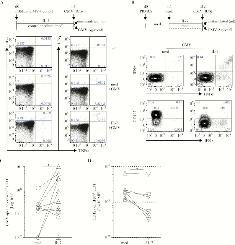
Interleukin (IL)-7 supports the accumulation of antiviral cytomegalovirus (CMV)-specific T cells. (A) Freshly derived peripheral blood mononuclear cells (PBMCs) from CMV+ donors were analyzed for inflammatory cytokine release after CMV-lysate intracellular cytokine staining (ICS) assay after a 7-day culture in the absence (d7, control medium [med], dashed black line) or presence of human recombinant IL-7 (d7, IL-7, black line). Left and right dot plots show the levels of IL-2 and tumor necrosis factor (TNF)α or interferon (IFN)γ and TNFα in gated CD4 T cells, respectively. Background levels of cytokine secretion were typically measured in unstimulated controls (nil). (B) Freshly isolated PBMCs from CMV+ donors were rested for 5 days in plain medium (dashed black line) before a 7-day culture in the absence (d12, med, dashed black line) or presence of human recombinant IL-7 (d12, IL-7, black line). At day 12 (d12), CD4+ T cells were analyzed for IFNγ and TNFα release (top row) alongside expression of CD127 (bottom row), after CMV-lysate ICS assay. Cytomegalovirus-specific IFNγ+ CD4+ T cells show high CD127 expression in the resting cultures, whereas they downregulated CD127 expression upon exposure to IL-7. Background levels of cytokine secretion were typically measured in ICS unstimulated (nil) controls. A representative of >10 independent experiments is shown. (C) After subtraction of individual background levels of IFNγ+ and TNFα+ and IFNγ+TNFα+ CD4 T cells detected in unstimulated controls (nil), the percentage of CMV-specific cytokine+ (IFNγ+ and TNFα+) CD4 T cells was evaluated in 10 independent CMV+ donors in IL-7 (IL-7, open triangles) compared with control medium ([med], open circles) cultures. The graph shows a statistically significant (paired Wilcoxon test, *P = .03) increase of the frequency (Log10) of CMV-specific CD4 T cells after IL-7 culture. (D) CD127 expression (Log10 mean fluorescence intensity [MFI]) is significantly downregulated in CMV-specific IFNγ+CD4+ T cells exposed to IL-7 (IL-7) compared with control medium (med) at d12 in 6 biologically independent replicates (paired Wilcoxon test, *P = .03).

