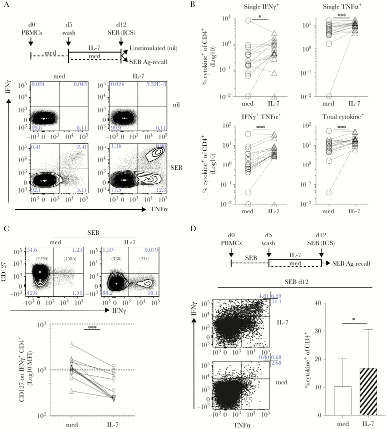
Figure 4.
Interleukin (IL)-7 supports superantigen-specific responses. (A) Freshly isolated peripheral blood mononuclear cells (PBMCs) were rested for 5 days in plain medium (dashed black line) then incubated for 1 week in IL-7 or control medium ([med] respectively IL-7, black line and med, dashed black line). At day (d)12, cells were stimulated with Staphylococcus aureus enterotoxin B (SEB) followed by an intracellular cytokine staining (ICS) assay to test antigen (Ag)-specific tumor necrosis factor (TNF)α and interferon (IFN)γ release, compared with unstimulated controls (nil). Dot plots show that the frequency of SEB-specific CD4+ T cells producing TNFα and/or IFNγ increased upon exposure to IL-7 in 1 representative donor. (B) After subtraction of individual background levels of IFNγ+ and TNFα+ and IFNγ+TNFα+ CD4 T cells detected in unstimulated controls (nil), the percentage of SEB-specific cytokine+ (IFNγ+, TNFα+, IFNγ+TNFα+ and total) CD4 T cells was evaluated in 17 independent donors in IL-7 (IL-7, open triangles) compared with control medium (med, open circles) cultures. The graphs show a statistically significant increase of the frequency (Log10) of SEB-specific cytokine+ CD4 T cells after IL-7 culture. All tests are paired Wilcoxon tests with the exception of the single TNFα+ analysis (paired t test): *, P ≤ .05; **, P ≤ .005; ***, P ≤ .0005. (C) At d12, cells cultured as in (A) were tested for CD127 expression in parallel to cytokine release in SEB-specific ICS assay. CD127 expression (Log10 mean fluorescence intensity [MFI]) is significantly decreased in SEB-specific IFNγ+ CD4+ T cells exposed to IL-7 (IL-7, downward triangles) compared with control medium (med, upward triangles) in 12 independent biological replicates (paired Wilcoxon test; ***, P = .0005). (D) Freshly isolated PBMCs (d0) were stimulated with the SEB superantigen (dotted black line) for 5 days (d0–5). At d5, cells were washed and incubated for 1 week in IL-7 (IL-7, black line; d5–12) or control medium (med, dashed black line; d5–12). At d12, cells were stimulated with SEB (overnight) to test Ag-specific TNFα and IFNγ release, by ICS. Dot plots show that the frequency of SEB-specific CD4+ T cells producing TNFα and/or IFNγ increased upon exposure to IL-7 (IL-7 d5–12, top row). The graph on the right shows data from the same cultures derived from 6 independent biological replicates. Statistically significant accumulation of SEB-specific, cytokine+ CD4+ T cells was evaluated using a Wilcoxon matched-pairs signed-ranked test (P = .03).

