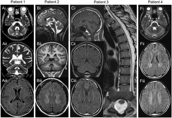
Figure 2.
MRI findings. Brain or Spinal MRI of (A) Patient 1 at age 57, (B) Patient 2 at age 18, (C–E) Patient 3 at age 26 and (F). Axial and coronal T2-weighted images show mild cerebellar atrophy and enlarged fourth ventricle (Ai, ii and Fi). Sagittal T2-weighted and coronal T1-weighted images show mild cerebellar atrophy (Bi and ii). Sagittal T1-weighted and coronal fluid-attenuated inversion recovery (FLAIR) images show mild cerebellar atrophy (Ci and ii). FLAIR image of Patient 2 (Biii) and Patient 4 (Fii and iii) shows mild bilateral hyperintensity in the periventricular white matter, but not of Patients 1 (Aiii) and 3 (Ciii). Sagittal and axial T2-weighted images of the spine of Patient 3 show spinal cord atrophy with no intrinsic cord signal abnormality (D and E).

