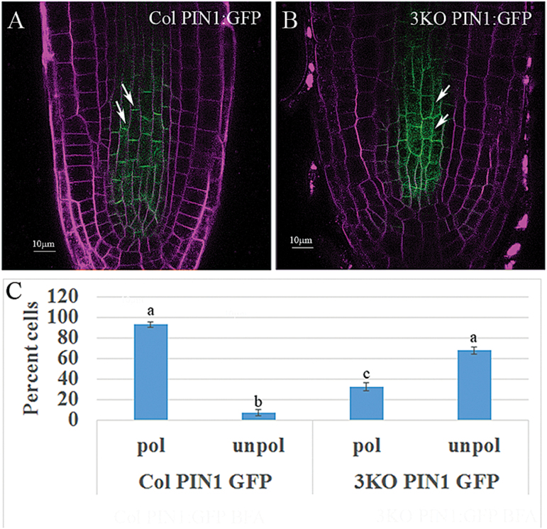
Fig. 4.
Loss of polar localization of PIN1:GFP in the stele cells of 3KO plants. PIN1:GFP was expressed in 3KO plants and its distribution in stele cells was compared with Columbia plants also expressing this reporter. (A) Columbia PIN1:GFP root tip; arrows mark two cells with typical polarized PIN1:GFP localization. (B) 3KO PIN1:GFP; arrows point to cells exemplifying depolarized PIN1:GFP localization. (C) Quantitative analysis of the proportion of cells (%) showing depolarized PIN1:GFP, evaluated by Scheffe analysis. Letters denote samples showing statistically insignificant (two ‘a’s) and statistically significant (‘a’ versus ‘b’ and ‘c’; ‘b’ versus ‘c’; P<0.05) differences. Scale bars=10 µm.

