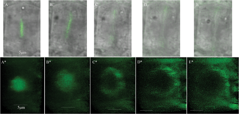
Fig. 5.
Time-lapse imaging of the myosin XI-K:YFP in 3KOR root meristem cells reveals dynamic disc-like structures that gradually accommodate ring-like shapes. Imaging has been done at 5 min intervals. (A–E) Myosin XI-K:YFP imaging in a single cell. (A*–E*) Corresponding tilted 3-D reconstructions of the myosin XI-K:YFP signal from each time interval performed using a ×63/1.2 objective and Z sectioning of 9 µm (total of 61 optic sections, every 0.15 µm with frame accumulation ×3 and resolution 512 × 512). The Leica Hyd detector and speed scan at 600 Hz were used. Scale bars=5 µm.

