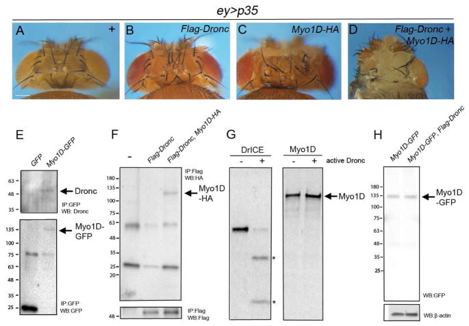
Figure 3. Genetic and physical interaction between Myo1D and Dronc.
(A–D) Head capsule phenotypes of the indicated genotypes. Scale bar: 200μm.
(E) Extracts from ey>hid,p35,GFP and ey>hid,p35;Myo1D-GFP eye/antennal imaginal discs were immunoprecipated with anti-GFP antibodies. Shown are immunoblots of Myo1D-GFP immunoprecipitates probed with anti-Dronc (SK11) (top) and anti-GFP antibodies (bottom). Arrows indicate Myo1D-GFP and Dronc.
(F) Extracts from ey-Gal4 (-), ey>Flag-Dronc and ey>Flag-Dronc,Myo1D-HA eye/antennal imaginal discs were immunoprecitated with anti-Flag antibodies. Shown are immunoblots probed with anti-HA antibodies to visualize Myo1D-HA (top, arrow) and with anti-Flag antibodies to reveal Flag-Dronc (bottom).
(G) Autoradiographs of in vitro cleavage assays of radio-labeled DrICE (positive control) and radio-labeled Myo1D by unlabeled active Dronc. Asterisks mark the cleavage products of DrICE.
(H) Immunoblot analysis of total extracts from ey>hid,p35;Myo1D-GFP eye/antennal imaginal discs using anti-GFP antibodies.

