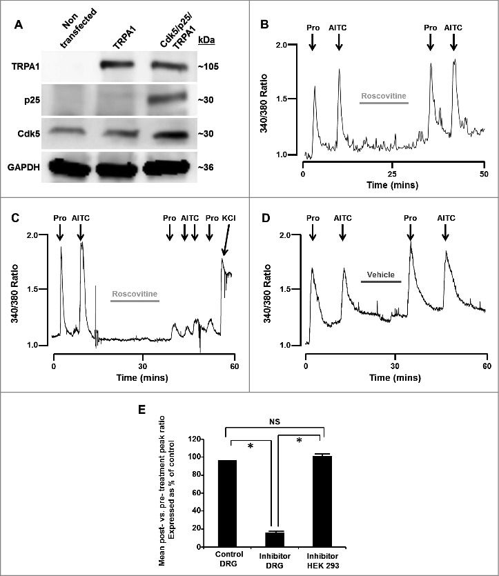
Figure 1.

(A) Representative western blot images depicting the presence or absence of TRPA1, p25 or Cdk5 in HEK293 cells that were either non-transfected or transfected with TRPA1 only or Cdk5, p25 and TRPA1. GAPDH was probed as a loading control. (B) Representative trace depicting [Ca2+]i transients induced by propofol and AITC (TRPA1 agonists) in the absence and presence of roscovitine (50 µM) in TRPA1/TRPV1-transfected HEK293 cells (which lack Cdk5 activity). Cells were exposed to propofol (Pro, 10 µM) and AITC (100 µM) for 60 seconds at the time points marked by arrows. (C) Representative trace depicting [Ca2+]i transients induced by propofol and AITC in the absence and presence of roscovitine (50 µM) in DRG neurons. Cells were exposed to potassium chloride (KCl, 50 mM) for 30 seconds to demonstrate cell viability after exposure to roscovitine. (D) Representative control trace depicting [Ca2+]i transients induced by propofol and AITC in the absence and presence of DMSO vehicle (0.1%) in DRG neurons. E) Summarized data for experiments represented in panels B-D expressed as % of control ± SEM. NS = Not significant. n = 8 separate experiments, p < 0.05.
