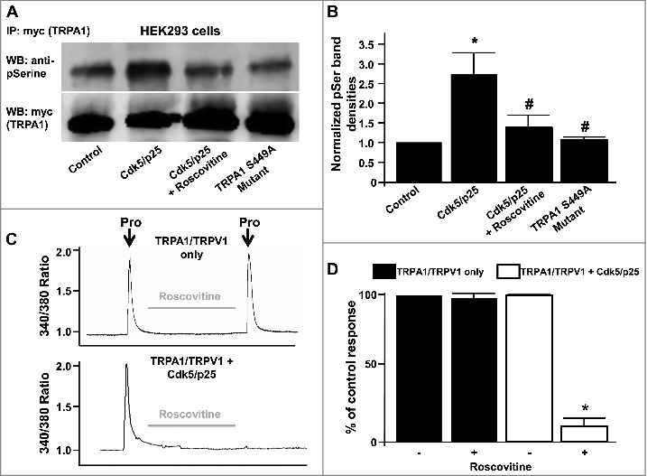
Figure 4.

(A) Representative western blot depicting phosphoserine immunoreactivity in HEK293 lysates from myc-tagged TRPA1 (myc-TRPA1) expressing cells co-transfected with either control RFP vector, Cdk5/p25 (in the presence or absence of roscovitine) or TRPA1 S449A mutant cDNAs. Lower blot shows myc-TRPA1 immunoreactivity in the same lanes after stripping and reprobing. (B) Summarized data for panel A, illustrating normalized TRPA1 phosphoserine immunoreactivities ± SEM. n = 6, p < 0.05. (C) Representative traces depicting effect of roscovitine on propofol-induced (Pro, 10 µM) transient increase in [Ca2+]i in HEK293 cells expressing TRPA1/TRPV1 only (top) or TRPA1/TRPV1 and Cdk5/p25 (bottom). (D) Summarized data for panel C expressed as percent of the control response normalized to 100% ± SEM. n = 8 separate experiments, p < 0.05.
