FIG 3.
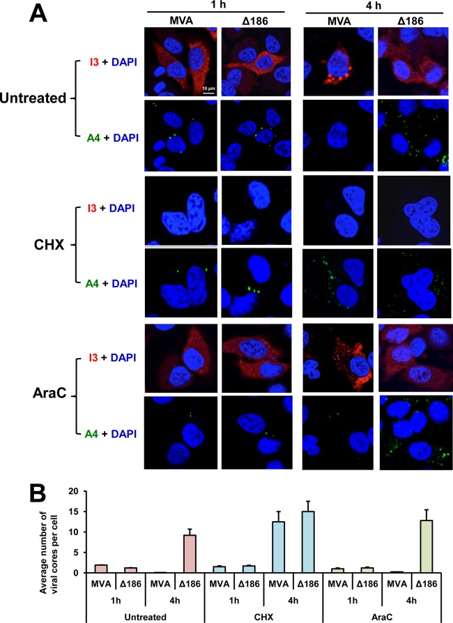
Deletion of MVA ORF 186 encoding the 68k-ank protein prevents viral genome uncoating. HeLa cells were untreated or treated with 100 μg/ml of cycloheximide (CHX) or 44 μg/ml of cytosine arabinoside (AraC) and infected with 5 PFU/cell of MVA or MVA-Δ186. After 1 h of adsorption at 4°C, the cells were washed and incubated at 37°C for 1 or 4 h. Cells were then fixed, permeabilized, and immunostained with mouse anti-I3 MAb and rabbit anti-A4 polyclonal antibody, followed by Alexa Fluor 568 anti-mouse IgG and Alexa Fluor 647 anti-rabbit IgG, respectively. Nuclei were stained blue with DAPI. (A) The subcellular localizations of I3 (red), A4 (green), and DAPI (blue) were determined by confocal microscopy, with the scale bar representing 10 μm. (B) Viral cores that stained with anti-A4 antibody and cells that stained with DAPI were enumerated from 40 to 120 cells, and the average numbers of viral cores per cell were plotted. Standard deviations were calculated from the numbers obtained in three random fields.

