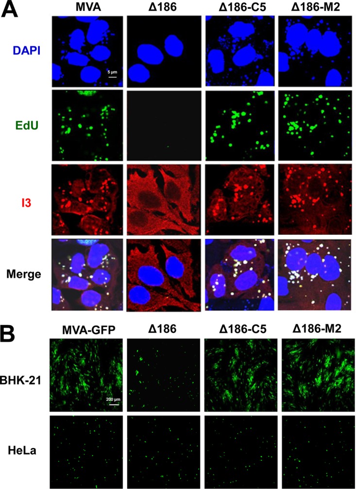
FIG 8.

C5 and M2 restore genome replication by MVA-Δ186. (A) HeLa cells were infected with 5 PFU/cell of MVA, MVA-Δ186, rMVA-Δ186-C5, or rMVA-Δ186-M2. At 3 h after infection, the cells were incubated with EdU (10 μM) and then fixed, permeabilized, and reacted with Alexa Fluor 647 azide. The I3 single-stranded DNA binding protein was visualized by staining with a MAb followed by an anti-mouse secondary antibody conjugated to Alexa Fluor 568. DAPI was used to stain total DNA. Images were collected with a confocal microscope. The scale bar represents 5 μm. (B) BHK-21 and HeLa cells were infected with 0.05 PFU/cell of the indicated viruses expressing GFP under the control of the P11 late promoter for 24 h. Virus spread was visualized by confocal microscopy. The presence of similar numbers of cells was confirmed by differential image contrast (DIC) microscopy (not shown). The scale bar represents 200 μm.
