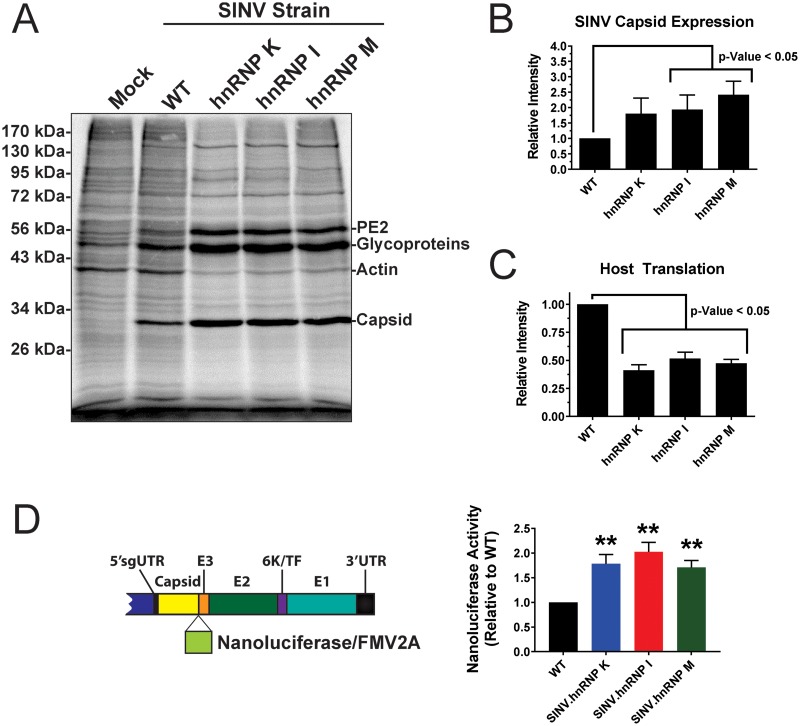
FIG 7.
Viral gene expression is altered following disruption of hnRNP-vRNA binding. (A) 293HEK cells were either mock treated or infected with wild-type parental virus or an individual hnRNP-vRNA interaction site mutant at an MOI of 10 PFU/cell, as indicated. At 10 h postinfection, the cells were pulsed for a period of 2 h with [35S]methionine and [35S]cysteine. After this period, the cells were harvested, and equal volumes of cell lysate were analyzed by SDS-PAGE and phosphorimaging. The data shown are representative of several independent biological replicates. (B) Densitometric quantification of the SINV capsid protein, with intensity relative to wild-type parental virus shown on the y axis. (C) Densitometric quantification of the host actin protein, with intensity relative to wild-type parental virus shown on the y axis. (D) Schematic diagram of the SINV nanoluciferase reporter used in this study. 293HEK cells were infected with either parental wild-type SINV or individual hnRNP-vRNA interaction site mutants/nanoluciferase reporters, and at 16 hpi, the level of nanoluciferase activity was quantified. All the quantitative data shown represent the means of three independent biological replicates, with the error bars representing standard deviations of the mean. Statistical significance was determined by Student's t test; **, P < 0.01.

