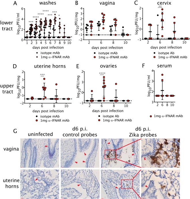
FIG 1.
Replication of ZIKV throughout the FRT after intravaginal infection. Five-week-old female C57BL/6 mice were injected subcutaneously (s.c.) with DMPA and inoculated intravaginally (ivag) with 104 PFU of ZIKV. One day prior to infection, mice were treated intraperitoneally (i.p.) with 1 mg anti-IFNAR1 mAb or an isotype control. (A) Vaginal washes were collected daily after infection, and replicating ZIKV was measured by plaque assay. Day 1, n = 15 to 17; day 2, n = 22 to 25; day 3, n = 13; days 4 to 6, n = 12; day 7, n = 7; day 8, n = 4; days 9 to 10, n = 2. (B to E) At days 2, 6, 8, and 10 p.i., mice were sacrificed, and the vagina (B), cervix (C), uterine horns (D), and ovaries (E) were harvested for virus titration by plaque assay. (F) Viremia was measured by plaque assay. Isotype, n = 5 to 6; anti-IFNAR1, n = 4 to 10. Data in panels A to F were pooled from three independent experiments. Statistical significance was measured by two-way ANOVA with Bonferroni's post hoc test. *, P < 0.05; **, P < 0.01; ***, P < 0.001; ****, P < 0.0001. Horizontal bars indicate the means, error bars show SD, and dashed lines show the limit of detection. (G) Representative images of ZIKV RNA detected by in situ hybridization in uninfected or ZIKV-infected mice at day 6 p.i. using ZIKV-specific or negative-control probes. The first three columns on the left show ×40 magnification, and the rightmost images show the inset outlined in red at ×63. The top row shows images for the vagina, and the bottom row shows images for the uterine horns. Arrows point to the lumenal edge of epithelium. All images are counterstained with hematoxylin. Scale bars, 50 μm in 40× images and 20 μm in 63× images. Data are representative of 3 animals per group.

