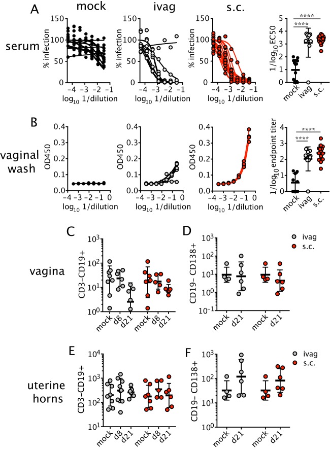
FIG 4.
Virus-specific antibodies are present in circulation and the vagina after ZIKV infection. Mice were infected ivag or s.c. with ZIKV after anti-IFNAR1 mAb injection as described in the legend for Fig. 2A. At 3 weeks p.i., neutralizing antibody was measured in the serum by FRNT (n = 10 to 21) (A), and E protein-specific IgG in the vaginal lumen was measured by ELISA (n = 9 to 14) (B). Graphs on the left were used to generate EC50s and endpoint titer dilutions. Each curve represents data from an individual animal. Total B cells (C and E) and antibody-secreting cells (D and F) were measured in the vagina (C and D) and uterine horns (E and F) on the indicated days. For CD19+ cells and both tissues and infection routes, n = 5 to 8. For CD138+ cells and both tissues and infection routes, n = 4 to 6. Horizonal bars show the means, and error bars show SD. Statistical significance was determined by one-way ANOVA with Bonferroni's post hoc test for panels A and B and by two-way ANOVA with Bonferroni's post hoc test on log-transformed data for panels C to F. Data were pooled from four independent experiments. ****, P < 0.0001.

