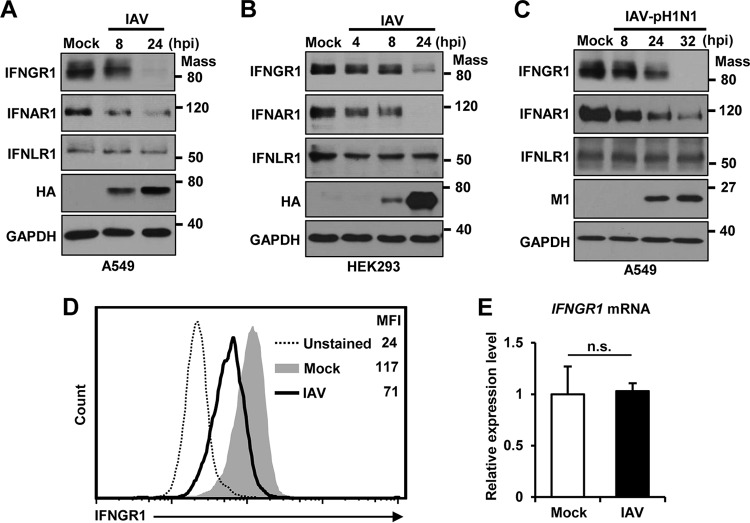
FIG 1.
IAV infection reduces the level of IFNGR1 protein. (A and B) A549 cells (A) and HEK293 cells (B) were infected with IAV at an MOI of 1. The levels of IFNGR1, IFNAR1, IFNLR1, and viral HA were analyzed by Western blotting at the indicated time points postinfection. The levels of GAPDH were used as an internal loading control. (C) A549 cells were infected with pandemic IAV-pH1N1 at an MOI of 1. Cells were harvested at the indicated time points, and the levels of IFNGR1, IFNAR1, IFNLR1, viral M1, and GAPDH were detected by Western blotting. (D) HEK293 cells were mock infected (Mock) or infected with IAV at an MOI of 1 (IAV). At 24 hpi, cells were left unstained or incubated with antibody against IFNGR1, followed by staining with FITC-conjugated secondary antibody. The surface expression levels of IFNGR1 were assessed by flow cytometric analysis. The MFI of each sample is shown. The experiment was repeated three times. (E) HEK293 cells were left uninfected (Mock) or infected with IAV at an MOI of 1. The relative mRNA levels of IFNGR1 were analyzed by real-time qPCR at 24 hpi. The data represent means and SD calculated from three reactions per sample. The experiment was independently repeated twice with similar results. n.s., not significant. Mass, molecular mass (kilodaltons).

