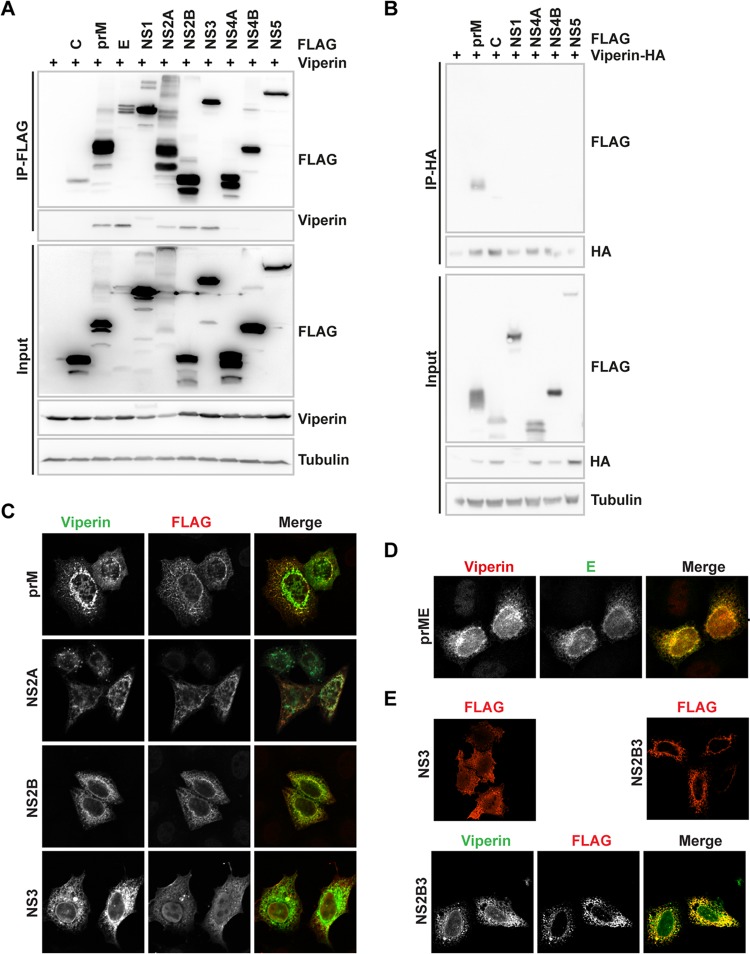
FIG 1.
Viperin interacts and colocalizes with TBEV prM, E, NS2A, NS2B, and NS3. (A) HEK293T cells were transiently transfected with plasmids encoding wild-type viperin and FLAG-tagged TBEV proteins and were subjected to anti-FLAG immunoprecipitation. Whole-cell lysates (input) and immunoprecipitated (IP) proteins were analyzed by immunoblotting using anti-FLAG and anti-viperin antibodies, including input levels of tubulin as a loading control. (B) HEK293T cells were transiently transfected with plasmids encoding HA-tagged viperin and FLAG-tagged TBEV prM, C, NS1, NS4A, NS4B, and NS5 and were subjected to anti-HA immunoprecipitation. Whole-cell lysates (input) and immunoprecipitated (IP) proteins were analyzed by immunoblotting using anti-FLAG and anti-HA antibodies, including input levels of tubulin as a loading control. (C and D) Immunofluorescence analysis of HeLa cells transiently transfected with plasmids expressing FLAG-tagged TBEV prM, NS2A, NS2B, and NS3 along with viperin (C) or prME with viperin (D). Proteins were detected using anti-FLAG, anti-viperin, and anti-E antibodies. (E) HeLa cells were transiently transfected with plasmids expressing FLAG-tagged TBEV NS3, NS2B3, or NS2B3 together with viperin. Representative images and blots are shown.

