FIG 1.
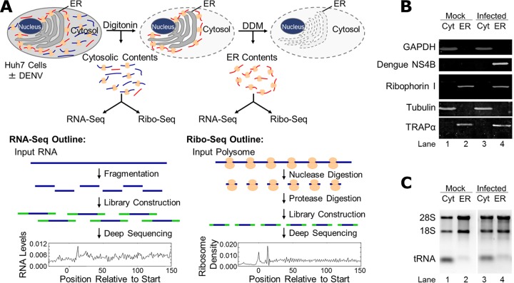
Experimental schematic and validation of cell fractionation protocol. (A) Schematic of the experimental approach. Mock- or DENV-infected Huh7 cells were fractionated by a sequential detergent extraction protocol where cell cultures are first treated with digitonin-supplemented buffers to release the cytosolic contents followed by a subsequent treatment with DDM-supplemented buffers to release the ER-associated contents. Total RNA was isolated from each fraction and analyzed by RNA-seq to assess gene expression. In parallel, polysomes in each fraction were nuclease digested, and ribosome footprints were isolated and analyzed by Ribo-seq. (B) Immunoblot analysis of the distributions of cytosolic (GAPDH and tubulin) and ER-resident membrane (ribophorin I and TRAPα) proteins in the cytosol (Cyt) and ER fractions of mock-infected cells and following 40 h of DENV infection (MOI of 10). (C) Ribosome and tRNA distributions in the two subcellular fractions were determined by isolation of total RNA, separation by agarose gel electrophoresis, and visualization with SYBR green staining. 18S, 28S, and tRNA components are indicated.

