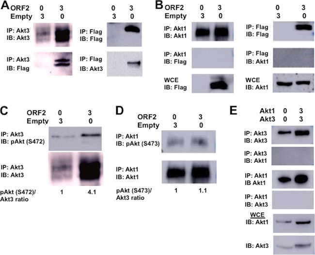
FIG 10.
Analysis of the effect ORF2 has on Akt3 and Akt1. For all studies described below, Neuro-2A cells were transfected with the designated amount of plasmids (μg DNA), and whole-cell extract (WCE) was collected at 48 h after transfection. (A) IP was performed with an Akt3 antibody (4059; Cell Signaling Technology) using WCE (400 μg protein). Immunoprecipitated proteins bound to magnetic protein A beads (S1425S; Invitrogen) were washed extensively, suspended in SDS-PAGE buffer, and separated in a 10% SDS-PAGE gel. After proteins were transferred to a PVDF membrane, the immunoblot (IB) was probed with the Akt3 antibody or anti-Flag monoclonal antibody (F1804; Sigma). (Right) IP was performed with the Flag antibody to IP ORF2 using total cell lysate (400 μg protein). Immunoprecipitated proteins were processed as described here and separated in a 10% SDS-PAGE gel, and the IB was probed with the Flag antibody or Akt3 antibody. (B) IP was performed with the Akt1 antibody (2938; Cell Signaling Technology) or Flag antibody using total cell lysate (400 μg protein). Immunoprecipitated proteins were analyzed by Western blotting and then probed with an Akt1 or anti-Flag monoclonal antibody. Western blot of WCE (50 μg protein) was probed with Flag or Akt1 antibody. (C) IP was performed with the Akt3 antibody, the precipitated proteins separated by 10% SDS-PAGE, and protein transferred to a membrane. The Western blot was then probed with the Akt antibody that recognizes phosphorylated Akt at serine 472 (pSer473; 9271; Cell Signaling Technology) or Akt3 antibody. ORF2 in WCE was detected using the Flag antibody. This was the same image as that shown in panel A, because these studies were performed in the same experiment. Levels of pSer472 Akt3 were quantified using ImageJ. (D) IP was performed with the Akt1 antibody using total cell lysate (400 μg protein). Immunoprecipitated proteins were analyzed in Western blots and then probed with the Akt antibody that recognizes phosphorylated Akt at serine 473 or anti-Akt1 antibody (2938; Cell Signaling Technology). WCE (50 μg protein) was probed with the anti-Flag monoclonal antibody to detect ORF2 (F1804; Sigma). Levels of phosphorylated serine 473 on Akt1 were quantified. (E) IP was performed with the Akt1 or Akt3 antibody using total cell lysate (400 μg protein). As described for earlier panels, immunoprecipitated proteins were examined for Akt1 or Akt3. Akt1 or Akt3 in 50 μg WCE was identified by Western blot analysis as a control. The results shown are representative of 3 independent studies.

