1.
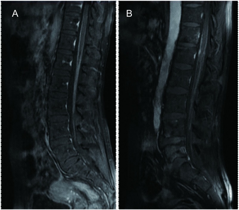
No.3(A)及No.2(B)患者T1压脂腰椎增强MRI表现。腰椎增强MRI显示软脊膜不均匀增厚伴有线样强化。
Sagittal sections of a contrast-enhanced fat-saturated T1-weighted magnetic resonance imaging (MRI) study of the lumbar spinal cord of patient No.2 (B) and patient No.3 (A).Contrast-enhanced MRI of lumbar spine showed diffuse abnormal enhancement of pial lining of spinal cord.

