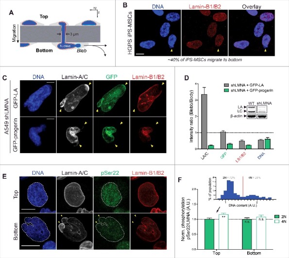
Figure 3.

Farnesylated progerin, as with lamin-B1/B2, is depleted from nuclear blebs following constricted migration through narrow pores. (A) Cartoon illustrating transwell migration of cells. Pore diameter = 3 μm; Polystyrene membrane thickness ∼10 μm. (B) Confocal images of HGPS iPS-MSCs that migrate to the bottom of narrow 3 μm pores (∼40% of seeded cells migrate to bottom in 48h) show typical nuclear blebs with lamin-B1/2 depletion. Scale bar = 10 μm. (C) Representative images of nuclei exhibiting characteristic blebs following constricted migration. Lamin-A/C is enriched in sites of nuclear blebs (as seen by immunofluorescence and GFP signal), but GFP-progerin is depleted from blebs, as are the farnesylated lamins-B1 and B2. Scale bar = 5μm. (D) Quantitation of nuclear bleb/body fluorescence intensity ratio. Inset: immunoblot of WT and shLMNA cells showing residual LA and LC. (E) Confocal images of WT A549 cells at the top & bottom of the transwell membrane. Phosphorylated lamin-A/C (‘pSer22’) is seen in the nucleoplasm of interphase nuclei. Cells that migrate to the bottom show nuclear blebs with lamin-A/C enrichment and lamin-B1/B2 depletion (yellow arrowheads). Scale bar = 10 μm. (F) While lamin-A/C is phosphorylated ∼10-15% higher in ‘4N’ vs ‘2N’ cells (inset), normalized phosphorylation (as ‘pSer22/LMNA’) does not change significantly before and after migration.
