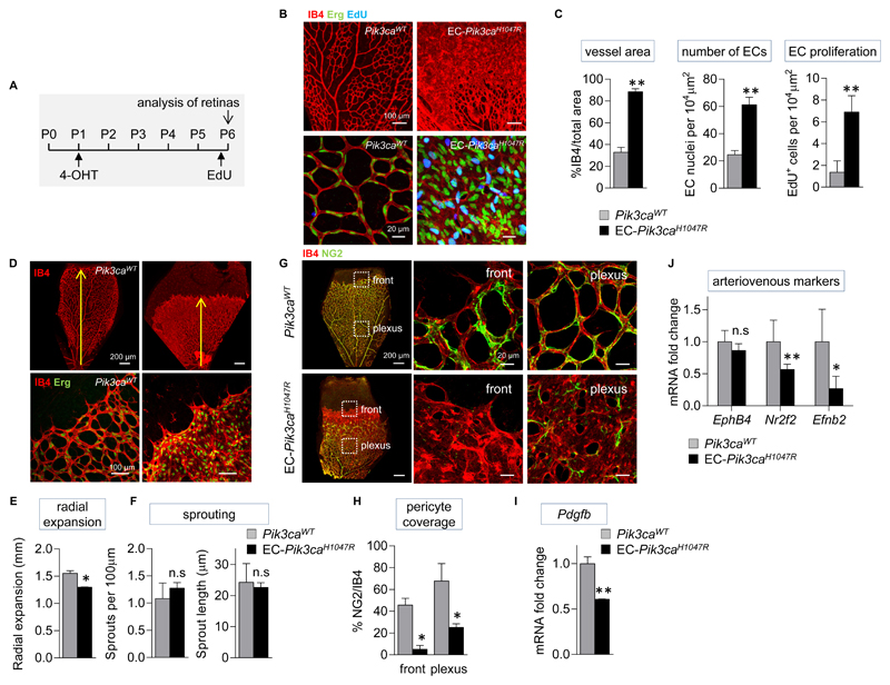
Figure 3. Endothelial activation of Pik3ca promotes hyperproliferation in ECs and impairs pericyte coverage.
(A) Schematic of the 4-OHT and EdU administration regime used. (B) Representative flat-mounted Pik3caWT and EC-Pik3caH1047R P6 retinas stained with IB4 (red, revealing ECs) and antibody to the Erg transcription factor (nuclear marker of ECs; green) and labelled with EdU (blue). (C) Quantitative analysis of the retina vessel area (assessed by IB4 staining), EC numbers (assessed by staining for Erg), and number of proliferating ECs (cells positive for both EdU and Erg). Data represent mean ± SEM. **p ≤ 0.01 (Mann-Whitney U test). n=6/genotype. (D) Representative flat-mounted control and EC-Pik3caH1047R P6 retinas stained with IB4 and antibody to the Erg transcription factor. (E) Quantification of the radial expansion of vasculature in retinas. Data represent mean ± SEM. *p < 0.05 (Mann-Whitney U test). n=6/genotype. (F) Quantification of the number of sprouts at the vascular front per unit length, and the length of sprouts. Data represent mean ± SEM. n.s., not significant, p > 0.05 (Mann-Whitney U test). n=6/genotype. (G) Flat-mounted Pik3caWT and EC-Pik3caH1047R retinas showing vasculature (IB4; red) and pericytes (stained for NG2, a membrane proteoglycan found in pericytes; green). Right, higher magnification of highlighted sections. (H) Quantification of pericyte coverage in the vasculature of retinas (assessed by % of NG2 staining relative to IB4 staining). Data represent mean ± SEM. *p < 0.05 (Mann-Whitney U test). n=6/genotype. (I) Pdgfb mRNA expression in EC-Pik3caH1047R P6 retinas. Data represent mean ± SEM. **p < 0.01 (Mann-Whitney U test). n=5/genotype. (J) Ephb4, Nr2f2, and Efnb2 mRNA expression in EC-Pik3caH1047R P6 retinas. Data represent mean ± SEM. n.s., not significant, p > 0.05, *p < 0.05, **p < 0.01 (Mann-Whitney U test). n=5/genotype.

