3.
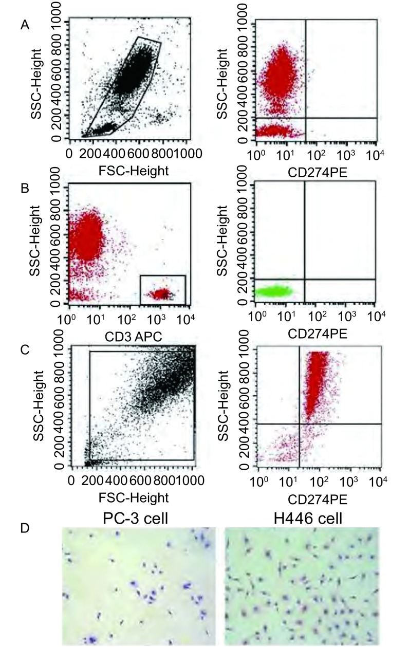
PD-L1在SCLC外周血和H446细胞中的表达及分布。A:左图,外周血单个核细胞群(R1门);右图,R1门中PD-L1水平;B:左图,CD3+ T细胞群(R2门);右图,R2门中PD-L1水平;C:PD-L1在H446表达(FACS法);D:PD-L1在H446细胞中表达(ICC法)。PC-3细胞为阴性对照。FACS:流式细胞术;ICC:免疫细胞化学法。
Distribution and expression of PD-L1 in peripheral blood of SCLC patients and H446 cells. A: Left, mononuclear cells in peripheral blood (Gate R1); Right, Level of PD-L1 in Gate R1; B: Left, CD3+ T cells (Gate R2); Right, PD-L1 in Gate R2; C: The expression of PD-L1 in H446 cells (FACS); D: PD-L1 expression in H446 cells (ICC) with PC-3 cell as negative control. FACS: flow cytometry; ICC: immunocytochemical method.

