2.
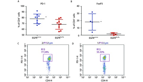
基因组小鼠肿瘤酶消化悬液细胞分析。EGFRflx/flx相对于EGFRwt/wt组,PD-1阳性CD4+ T细胞比例降低(A),FoxP3阳性CD4+ T细胞比例也明显下降(B)。EGFRwt/wt组样本PD-1阳性CD4+T为77.34%(C),同源EGFRflx/flx样本PD-1阳性CD4+ T为55.33%(D)。*P<0.05。
FACS analysis the tumor cell suspensions in the genetic model. EGFRflx/flx keratinocytes formed tumors vs the EGFRwt/wt, the proportion of PD-1 positive CD4+ T cells decreased (A), FoxP3 positive CD4+ T cell ratio was also significantly decreased (B). A sample from EGFRwt/wt group, the PD-1 positive CD4+ T ratio was 77.34% (C), the ratio of EGFRflx/flx sample PD-1 positive CD4+ T was 55.33% (D). *P < 0.05.

