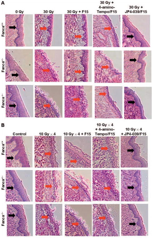
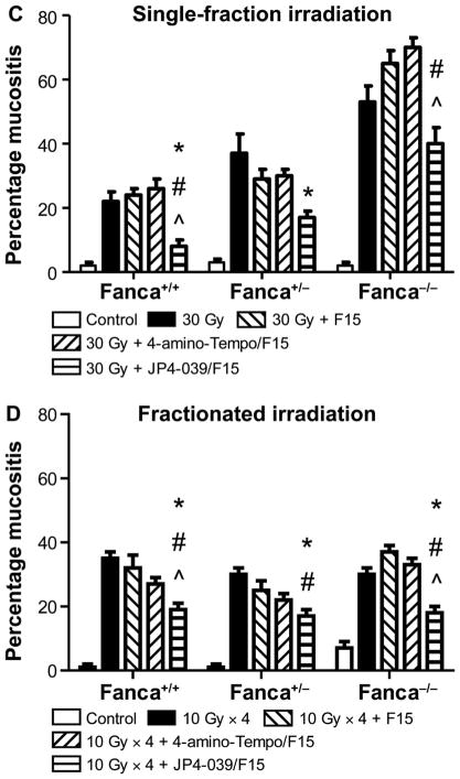
FIG. 5.
Histopathologic appearance of oral cavity tissue after single-fraction or fractionated head and neck irradiation of Fanca−/− mice and amelioration by JP4-039/F15 of radiation damage. Groups of mice (Fanca−/−, Fanca+/− and Fanca+/+; n =5 per group) were head and neck irradiated with a single 30 Gy fraction (panels A and C) or four successive days of 10 Gy (panels B and D) and all evaluated at 5 days after the final fraction. Panels A and B: H&E staining for single-fraction and fractionated irradiation groups, respectively. Black arrows indicate normal epithelium; red ← arrows indicate damaged/ulcerated epithelium. Panels C and D: Damage quantitation after single-fraction and fractionated irradiation, respectively. In panel C, for Fanca+/+, Fanca+/− and Fanca−/− values, respectively, when JP4-039 is compared to 30 Gy, *P < 0.0054, *P < 0.0075 and *P < 0.05042; when JP4-039 is compared to 30 Gy + F15, #P < 0.0005, #P < 0.2188 and #P < 0.0203; and when JP4-039 is compared to 30 Gy + 4-amino-Tempo/F15, ^P < 0.0002, ^P < 0.1435 and ^P < 0.0045. For Fanca+/+, Fanca+/− and Fanca−/− values in panel D, when JP4-039 is compared to 30 Gy, *P < 0.0001, *P < 0.0001 and *P =0.0002; when JP4-039 is compared to 30 Gy + F15, #P = 0.0006, #P = 0.0082 and #P < 0.0001; and when JP4-039 is compared to 30 Gy + 4-amino-Tempo/F15, ^P =0.0065, ^P =0.1015 and ^P < 0.0001.


