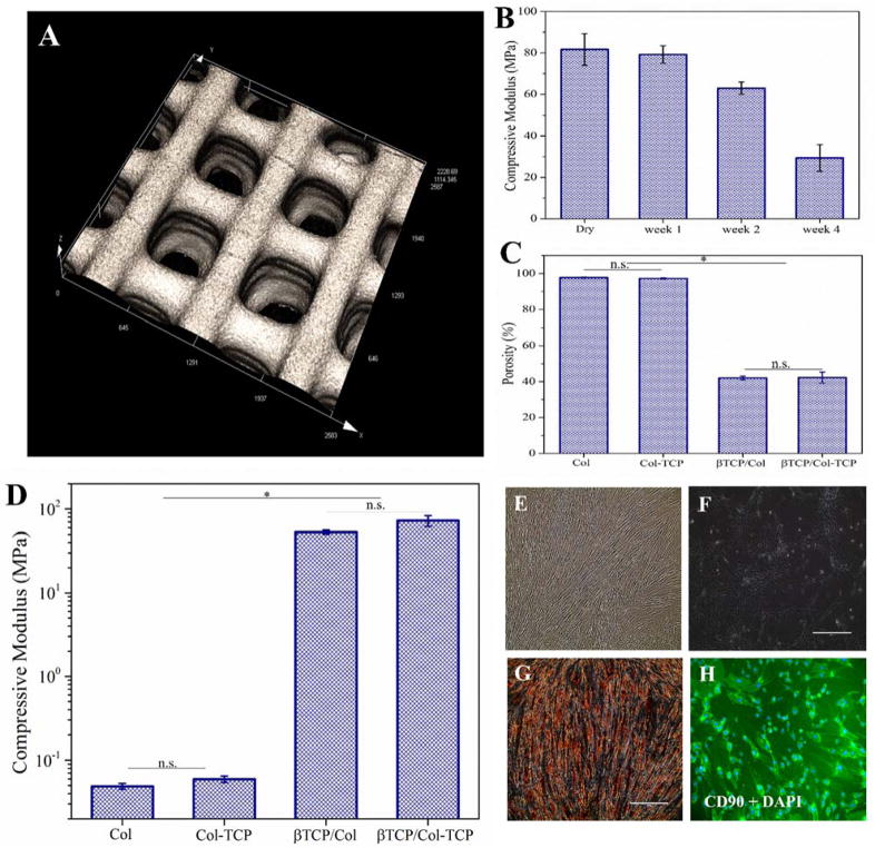
Figure 4.
(A) 3D laser scanning micrographs of the 3D-printed β-TCP scaffold (The scale bar is 500 μm). Porosity of Collagen and Collagen-TCP matrices compared with β-TCP/Col and β-TCP/Col-TCP hybrid constructs. (C) Compressive modulus of 3D-printed β-TCP scaffolds in dry state and after 1, 2, and 4 weeks immersion in phosphate-buffered saline. (D) Compressive modulus of Collagen and Collagen-β. TCP matrices compared with β-TCP/Col and β-TCP/Col-TCP hybrid constructs (E,F) optical image of 2D cultured DPCs. (G) Alizarin Red S staining of 2D cultured DPCs after three weeks osteogenic differentiation (scale bar is 400 μm). (H) Immunofluorescence images of 2D cultured DPCs using surface marker of CD90 and DAPI (scale bar is 400 μm).

