FIGURE 1.

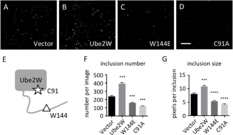
Ube2W alters Httex1Q103-GFP inclusion formation in HEK293 cells
A-D. In HEK293 cells, Httex1Q103 inclusions were visualized by GFP fluorescence 48 hrs after transfection.
Httex1Q103-GFP was co-expressed with vector alone (A), Ube2W (B), Ube2W-W144E (C) or Ube2W-C91A (D). Scale bar=30 μm.
E. Schematic of Ube2W structure illustrates relative positions of C91 (the active site cysteine) and W144 (needed for substrate binding).
F,G. Httex1Q103 inclusion number (F) and size (G) from all four groups were plotted. Graphs show means +/− SEM; ***, p<0.001; ****, p<0.0001. n=8 images(F), n>34 inclusions (G).
